Veterinaria (Montevideo), 2026 62(226), https://doi.org/10.29155/VET.62.226.6
Artículo original
Epigallocatechin-3-gallate (EGCG) improves in vitro maturation of sheep oocytes
El epigalocatequina-3-galato (EGCG) mejora la maduración in vitro de los ovocitos de oveja
Epigalocatequina-3-galato (EGCG) melhora a maturação in vitro de oócitos ovinos
Andreza Mayara Carneiro Lima1 https://orcid.org/0000-0001-6192-5937
Luana Keally Pimentel de Oliveira1 https://orcid.org/0000-0001-6440-5515
Érika Karoline de Oliveira Aureliano1 https://orcid.org/0009-0003-3875-2494
Damaris Raquel Pires dos Santos1 https://orcid.org/0009-0005-5269-7983
Bruna Moura da Silva1 https://orcid.org/0000-0003-2832-6689
Luanna Mendes Souza Rodrigues2 https://orcid.org/0009-0002-8994-6339
Gabriela Cristina da Silva Santos2 https://orcid.org/0000-0003-0876-6054
Rafael Soares dos Anjos1 https://orcid.org/0000-0002-5979-5229
Beatriz Cavalcanti de Freitas1 https://orcid.org/0009-0002-1073-1555
Neilton de Lima Oliveira Junior1 https://orcid.org/0009-0000-0974-3642
Brena Ferreira de Menezes1 https://orcid.org/0000-0002-3270-9490
Jennifer da Silva Veloso1 https://orcid.org/0009-0002-8863-1343
Pedro Humberto Félix de Souza3 https://orcid.org/0009-0004-0218-0743
Mabel Freitas Cordeiro1 https://orcid.org/0000-0003-1963-7311
Maria Helena Tavares de Matos2 https://orcid.org/0000-0001-6215-5292
Edilson Soares Lopes Júnior1 https://orcid.org/0000-0002-4790-8134
1 Laboratory of Physiology and Biotechnology of Animal Reproduction (LAFIBRA), Federal University of Vale do São Francisco, 56300-000, Campus of Agricultural Sciences, Petrolina-PE, Brazil. Corresponding author: andreza98carneiro@outlook.com
2 Nucleus of Biotechnology Applied to Ovarian Follicle Development, Federal University of Vale do São Francisco, 56300-000, Campus of Agricultural Sciences, Petrolina-PE, Brazil.
3Department of Technology and Social Sciences, State University of Bahia, 48900-000, Juazeiro-BA, Brazil.
Cómo citar este artículo:
Carneiro Lima, A. M., Pimentel de Oliveira, L. K, de Oliveira Aureliano, E. K., Pires dos Santos, D. R., Moura da Silva, B., Mendes Souza Rodrigues, L., da Silva Santos, G. C., Soares dos Anjos, R., Cavalcanti de Freitas, B., de Lima Oliveira Junior, N., Ferreira de Menezes, B., da Silva Veloso, J., Félix de Souza, P. H., Freitas Cordeiro, M., Tavares de Matos, M. H., & Soares Lopes Júnior, E. (2026). Epigallocatechin- 3-gallate (EGCG) improves in vitro maturation of sheep oocytes. Veterinaria (Montevideo), 62(226), e20266222606. https://doi.org/10.29155/VET.62.226.6
Abstract
This study evaluated the effect of epigallocatechin-3-gallate (EGCG) in ovine oocyte in vitro maturation (IVM) medium. Oocytes were assigned to IVM in four experimental groups: CON, composed of TCM-199 supplemented with 500 IU penicillin, 0.5 mg streptomycin, 1.25 µg amphotericin B, 0.2 mM sodium pyruvate, 10 % (v/v) FBS, and 15 IU/mL eCG; and the EGCG25, EGCG50, and EGCG100 groups, where they were matured in the same medium as the CON group, supplemented with 25 µM, 50 µM, and 100 µM EGCG, respectively. At the end of the maturation period, 1.053 oocytes were used for evaluation of cumulus cell expansion, chromatin configuration, and quantification of active mitochondria, reactive oxygen species (ROS), and glutathione (GSH) levels. 391 oocytes was used for in vitro fertilization (IVF) and in vitro embryo culture (IVC). The results showed that the EGCG50 group had higher rates of MII, active mitochondria, and GSH than the other groups, as well as a higher cleavage rate than the CON group. In conclusion, a concentration of 50 µM EGCG improves oocyte maturation rate and in vitro ovine embryo production.
Keywords: Antioxidante, Catechin, Sheep, IVP.
Resumen
Este estudio evaluó el efecto de la epigalocatequina-3-galato (EGCG) en el medio de maduración in vitro (MIV) de ovocitos ovinos. Los ovocitos fueron asignados al MIV en cuatro grupos experimentales: CON, compuesto por TCM-199 suplementado con 500 UI de penicilina, 0,5 mg de estreptomicina, 1,25 µg de anfotericina B, 0,2 mM de piruvato sódico, 10 % (v/v) de suero fetal bovino (SFB) y 15 UI/ml de EGCG; y los grupos EGCG25, EGCG50 y EGCG100, donde se maduraron en el mismo medio que el grupo CON, suplementado con 25 µM, 50 µM y 100 µM de EGCG, respectivamente. Al final del periodo de maduración, 1053 ovocitos se utilizaron para evaluar la expansión de las células del cúmulo, la configuración de la cromatina y la cuantificación de las mitocondrias activas, las especies reactivas de oxígeno (ROS) y los niveles de glutatión (GSH). Se utilizaron 391 ovocitos para la fertilización in vitro (FIV) y el cultivo de embriones in vitro (CIV). Los resultados mostraron que el grupo EGCG50 presentó mayores tasas de MII, mitocondrias activas y GSH que los demás grupos, así como una mayor tasa de clivaje que el grupo CON. En conclusión, una concentración de 50 µM de EGCG mejora la tasa de maduración de los ovocitos y la producción de embriones ovinos in vitro.
Palabras clave: Antioxidante, Catequina, Oveja, PIV.
Resumo
Este estudo avaliou o efeito da epigalocatequina-3-galato (EGCG) no meio de maturação in vitro (MIV) de oócitos ovinos. Os oócitos foram distribuídos em quatro grupos experimentais submetidos à MIV: CON, composto por meio TCM-199 suplementado com 500 UI de penicilina, 0,5 mg de estreptomicina, 1,25 µg de anfotericina B, 0,2 mM de piruvato de sódio, 10 % (v/v) de soro fetal bovino (SFB) e 15 UI/mL de eCG; e os grupos EGCG25, EGCG50 e EGCG100, nos quais os oócitos foram maturados no mesmo meio do grupo CON, suplementado com 25 µM, 50 µM e 100 µM de EGCG, respectivamente. Ao final do período de maturação, 1.053 oócitos foram utilizados para avaliação da expansão das células do cumulus, configuração da cromatina e quantificação de mitocôndrias ativas, espécies reativas de oxigênio (ERO) e níveis de glutationa (GSH). 391 oócitos foram utilizados para fecundação in vitro (FIV) e cultivo in vitro de embriões (CIV). Os resultados mostraram que o grupo EGCG50 apresentou maiores taxas de MII, mitocôndrias ativas e GSH do que os demais grupos, bem como uma taxa de clivagem superior à do grupo CON. Em conclusão, a concentração de 50 µM de EGCG melhora a taxa de maturação dos oócitos e a produção in vitro de embriões ovinos.
Palavras-chave: Antioxidante, Catequina, Ovelha, PIV.
Fecha de recibido: 15/04/2026
Fecha de aceptado: 31/08/2026
Introduction
Most research on in vitro embryo production (IVP) focuses on oocyte quality and the proper occurrence of nuclear and cytoplasmic maturation, processes essential to ensure developmental competence and fertilization capacity. One of the main challenges in in vitro maturation (IVM) of oocytes is the increased production of reactive oxygen species (ROS), related to high oxygen (O2) tensions, temperature changes, and light exposure (Du Plessis et al., 2008; Gaviria et al., 2019). Furthermore, supplementing culture media with high concentrations of biological fluids, such as fetal bovine serum (FBS) and bovine serum albumin (BSA), and metallic minerals, such as iron and copper, which induce ROS formation via Fenton and Haber-Weiss reactions, intensifies this effect, resulting in lipid peroxidation (Guerin, et al., 2001; Torres-Osorio et al., 2019). Excess ROS causes imbalance in the cellular redox system, causing oxidative stress (Mateo-Otero et al., 2021), altering cellular metabolism, morphology, or signaling pathways (Wang et al., 2019), and may even lead to cell death.
In this context, the addition of antioxidants to the IVM medium becomes necessary (Marí et al., 2020). Among the antioxidants, epigallocatechin-3-gallate (EGCG) stands out, characterized as a polyphenol belonging to the catechin subclass, abundantly present in green tea, representing approximately 50-80 % of total catechins (Alfke et al., 2021). Its antioxidant action occurs through the presence of the phenolic hydroxyl group, which provides hydrogen to the molecules and subsequently undergoes oxidation reactions to form a highly stable mixture, thus removing free radicals that are harmful to the cell (Zhuang et al., 2024). Furthermore, EGCG is also able to reduce EO damage by regulating the activity of antioxidant enzymes, such as catalase (CAT), superoxide dismutase (SOD) and glutathione (GSH) (Meng et al., 2008), and the expression of genes related to protein kinase B (Barberino et al., 2020).
EGCG improved the quality of murine oocytes subjected to in vitro aging, protecting against oxidative stress and mitigating the rate of oocyte aneuploidy (Zhang et al., 2023). This antioxidant protected porcine oocytes exposed to the pesticide Diazinon, reducing apoptosis, protecting meiotic progression and spindle assembly, and inhibiting mitochondrial and DNA damage (Gai et al., 2022). After in vitro culture of sheep ovarian tissue, EGCG maintained follicle survival and reduced apoptosis through the PI3K/Akt signaling pathway (Barberino et al., 2020). Furthermore, supplementation of IVM medium with EGCG resulted in improved oocyte quality from diabetic mice (Chao et al., 2023) and increased cumulus cell expansion and maturation rate of bovine oocytes by decreasing ROS (Huang et al., 2018).
However, there are no reports on the effect of EGCG during IVM of ovine oocytes. Therefore, the aim of this study was to evaluate the effect of including EGCG in the IVM medium of ovine oocytes on cumulus cell expansion, oocyte nuclear maturation stage, intracellular levels of active mitochondria, ROS, GSH, and cleavage rate.
Material and Methods
Ethical Aspects
The entire protocol and procedures involving animals were reviewed and ethically approved by the Animal Use Ethics Committee (CEUA) of the Federal University of Vale do São Francisco (UNIVASF), and are registered under the following number: 0001/300823.
Media and Reagents
Unless otherwise noted, all chemicals used in this study were purchased from Sigma Aldrich Chemical Co. (St. Louis, MO, USA).
Ovary collection, oocyte collection and selection
Ovine ovaries were obtained from a local slaughterhouse immediately after the slaughter of ewes without defined breed standard (SPRD). After 90 minutes of collection, the ovaries were transported to the laboratory for 30 minutes in a 0.9 % NaCl solution containing 0.05 g of pentabiotic/L, composed of basic streptomycin, basic dihydrostreptomycin, benzathine benzylpenicillin, procaine benzylpenicillin, and potassium benzylpenicillin, at a temperature of 33 °C to 34 °C in a thermal container. In the laboratory, the ovaries were washed three times in heated saline solution (33 °C to 34 °C), adjacent tissues were removed, and then kept in a water bath (MODEL 1102®, Fanem, Brazil) at 34 °C. To collect cumulus-oocyte complexes (COC), a vacuum pump (BV 003®, WTA, Brazil) was used, at a pressure of 18 mmHg, corresponding to 5 mL/min, coupled to an 18G intravenous catheter (SOLIDOR®, Lamedid Comercial & Serviços, Brazil) and a 50 mL Falcon tube containing the recovery medium, composed of TCM 199, supplemented with 25 mM HEPES, 50 IU/mL sodium heparin, 500 IU penicillin, 0.5 mg streptomycin and 1.5 µg amphotericin and 1 % (v/v) fetal bovine serum (FBS). After harvesting, the COC were poured into 90 × 15 mm petri dishes (Mylabor, Brazil), analyzed under a stereomicroscope (SMZ 645®, Nikon, Japan) and qualified according to Freitas and Melo (2010), with only Grades I and II being selected for in vitro maturation.
In vitro maturation of ovine oocytes
After classification, the selected COC were divided into four maturation groups: control group (CON), composed of TCM-199, supplemented with 500 IU/mL of penicillin, 0.5 mg of streptomycin, 1.25 µg of amphotericin B, 0.2 mM of sodium pyruvate, 10 % (v/v) of FBS and 15 IU/mL of equine chorionic gonadotropin (eCG); and the EGCG25, EGCG50, EGCG100 groups, based on the same IVM medium as the CON group, supplemented with 25 µM, 50 µM and 100 µM of EGCG, respectively. Approximately 15 COC were placed in 75 µL drops of IVM medium from each treatment group, under mineral oil in 60 × 15 mm petri dishes (Mylabor, Brazil) and incubated (CO2 Incubator®, Tecnal, Brazil) for 24 hours, at an average temperature of 38.5 °C, in a humidified atmosphere with 5 % CO2, in four replicates. EGCG concentrations were based on previous research that showed the antioxidant effect of this substance when used in the IVM medium of bovine oocytes (Huang et al. 2018).
Assessment of Cumulus Cell Expansion and Degree of Expansion
At the end of the maturation process, the oocytes (n = 671) were assessed for cumulus cell expansion, classifying them as “non-expanded” or “expanded”. Furthermore, the degree of cumulus cell expansion was estimated, classifying them as partially expanded or fully expanded, according to the methodology of Marei et al. (2010), using a stereomicroscope (SMZ 645®, Nikon, Japan).
Chromatin Configuration Assessment
After IVM, oocytes underwent chromatin assessment. To this end, oocytes (n = 280) with expanded cumulus cells were denuded by successive pipetting and subsequently incubated in drops of PBS with 10 mM Hoechst 33342 for 15 min at room temperature in the dark. Subsequently, the oocytes were evaluated under an epifluorescence microscope (Nikon E200, Tokyo, Japan) at 400× magnification. Nuclear chromatin configuration was classified according to the different meiotic stages: Germinal Vesicle (GV), Germinal Vesicle Breakdown (GVB), Metaphase I (MI), Telophase I (TI), and Metaphase II (MII).
Assessment of intracellular levels of GSH, ROS and active mitochondria
After analyzing the data from the previous stage and verifying the EGCG treatment group with the highest MII rate, the IVM was repeated as previously described, however, using only this group and the control group. The oocytes from these groups were intended for evaluation of intracellular levels of GSH, ROS and active mitochondria, as described by Andrade et al. (2024), with subtle modifications. Briefly, oocytes (n = 102) were denuded and incubated in the dark at 37 °C for 30 minutes in PBS with 100 mM MitoTracker Red (MitoTracker Red®, CMXRos, Molecular Probes, Melbourne, Victoria, Australia), 10 mM 2´,7´-dichlorodihydrofluorescein diacetate (H2DCFDA®, Invitrogen Corporation) and 10 mM 4-chloromethyl-6,8-difluoro-7-hydroxycoumarin (CellTracker Blue®; CMF2HC®; Invitrogen Corporation, Carlsbad, United States) to detect intracellular levels of active mitochondria, ROS and GSH through red (579 nm), green (460 nm) and blue (370 nm) fluorescence, respectively. The oocytes were observed under an epifluorescence microscope (Nikon E200, Tokyo, Japan) and the fluorescence intensity of the oocytes was analyzed using Image J software (version 1.47®; National Institutes of Health®, Bethesda, MD, USA), where regions of interest (ROIs) were selected, and the mean gray value was measured using the Analyze–Measure function.
In vitro fertilization of ovine oocytes
Another portion of the matured oocytes (n = 391) that were not evaluated in the previous steps proceeded to IVF, in which fresh semen collected through the artificial vagina method (Gonzales et al., 2002) from a ram with proven fertility was used. Immediately after collection, the semen was subjected to macroscopic analysis (color, odor, and volume) and microscopic analysis (mass motility and sperm concentration). Subsequently, 1 mL of semen was added to a discontinuous 45 %/90 % Percoll gradient. Motile sperm were recovered by centrifugation at 700G for 15 minutes. The precipitate was resuspended in 2 mL of IVF medium (SOF®, supplemented with 500 IU/mL penicillin, 0.5 mg/mL streptomycin, 1.25 µg/mL amphotericin B, and 10 µg/mL hypotaurine) and washed by centrifugation at 100G for 5 minutes. The new precipitate was resuspended in 2 mL of IVF medium and the final sperm concentration was adjusted to 1 × 10⁶ sperm/mL, in a total volume of 50 µL/drop of IVF medium. Finally, the selected sperm were allocated in 50 µL drops of IVF medium, under mineral oil, together with the mature oocytes, in groups of 15/drop, in a 60 × 15 mm petri dish (Mylabor, Brazil), in an incubator (CO2 Incubator®, Tecnal, Brazil), at 38.5 °C with a humidified atmosphere containing 5 % CO2, for a period of 20 hours, in four replicates.
In vitro culture of ovine embryos
After IVF, the presumptive zygotes (n = 391) were denuded by successive pipetting and subjected to in vitro culture for 48 hours in 75 μL drops of SOF medium supplemented with 3 mg/mL BSA, 500 IU penicillin, 0.5 mg streptomycin, and 1.25 µg amphotericin B, under mineral oil, in 60 × 15 mm petri dishes (Mylabor, Brazil), containing 15 structures/drop. The structures were cultured in an incubator (CO2 Incubator®, Tecnal, Brazil), at 38.5 °C with 5 % CO2, in four replicates. The number of cleaved embryos was recorded on the second day of culture with the aid of an inverted microscope (Inverted Trinocular Microscope 400X, Bel Engineering®, Italy).
Statistical Analysis
The variables of cumulus cell expansion, chromatin configuration, and cleavage-stage embryos were expressed as percentages and compared using the chi-square test in Epi Info software (Epi Info 7.2.5, Atlanta, GA, USA, 2021). Data on active mitochondria, ROS, and GSH levels were subjected to the D’Agostino Pearson normality test. Considering these nonparametric data, the Kruskal-Wallis test was used for comparisons. When main effects or interactions were significant, means were compared using the Student-Newman-Keuls test using the online statistical program SAS® OnDemand for Academics 9.04.01 (SAS 3.8 Enterprise Edition, USA, 2020). Differences were considered statistically significant when P < 0.05.
Results
Cumulus Cell Expansion
Regarding cumulus cell expansion, both the control group (CON) and the groups containing EGCG (EGCG25, EGCG50, EGCG100) showed cell expansion (Figure 1).
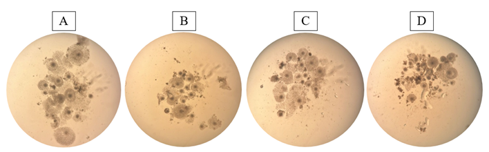
Figure 1: Expansion of cumulus cells of ovine oocytes after in vitro maturation in the absence of antioxidant supplementation (CON) (A) or in medium added with 25 µM (EGCG25) (B), 50 µM (EGCG50) (C) and 100 µM (EGCG100) (D) of EGCG
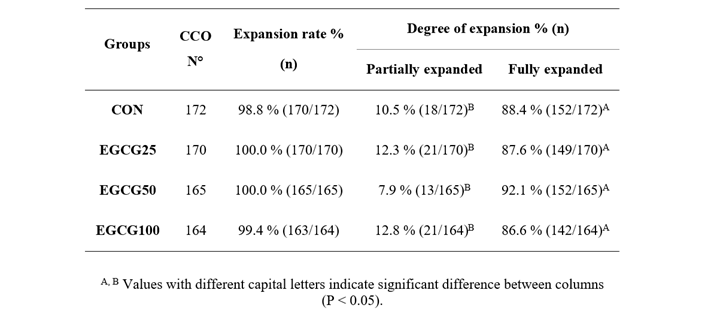
Regarding the degrees of expansion, there was no significant difference between the groups containing EGCG and the control group (CON) (Table 1; P > 0.05). However, when evaluated within each experimental group, it was possible to observe that, in all groups, the proportion of COCs with fully expanded cumulus cells was significantly higher (P-value: CON = 0.00; EGCG25 = 0.00; EGCG50 = 0.00; EGCG100 = 0.00) than the rate of COCs with partially expanded cells (Table 1).
Table 1. Degree of cumulus cell expansion (%) of ovine oocytes after in vitro maturation in the absence of antioxidant supplementation (CON) or in medium added with 25 µM (EGCG25), 50 µM (EGCG50) and 100 µM (EGCG100) of EGCG
Chromatin Configuration
Different meiotic stages were observed in the control group and in the groups containing different concentrations of EGCG (Figure 2).
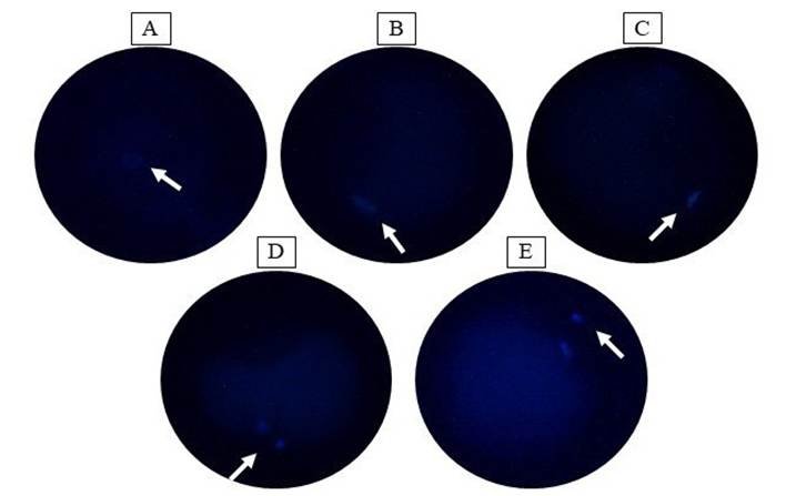
Figure 2:Chromatin configuration of ovine oocytes after in vitro maturation in the absence of antioxidant supplementation (CON) and in the groups supplemented with EGCG, via Hoechst 33342 probe. (A) Germinal vesicle in the CON group; (B) Germinal vesicle breakdown in the CON group; (C) Metaphase I in the EGCG25 group; (D) Telophase I in the EGCG100 group; (E) Metaphase II in the EGCG50 group
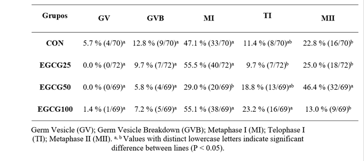
Regarding the GV and GVB meiotic stages, no significant difference was observed between the groups (Table 2; P > 0.05). However, for MI, the EGCG50 group presented a lower number of oocytes at this stage compared to CON (P-value = 0.04), EGCG25 (P-value = 0.00) and EGCG100 (P-value = 0.00) (Table 2). Furthermore, the EGCG100 group presented more TI compared to the EGCG25 group (P-value = 0.05), and the highest percentage of oocytes in MII was observed in the EGCG50 group when compared to CON (P-value = 0.00), EGCG25 (P-value = 0.01) and EGCG100 (P-value = 0.00) (Table 2).
Table 2. Chromatin configuration of ovine oocytes after in vitro maturation in the absence of antioxidant supplementation (CON) or in medium added with 25 µM (EGCG25), 50 µM (EGCG50) and 100 µM (EGCG100) of EGCG
Levels of active mitochondria, ROS, and GSH
Oocytes matured with 50 µM EGCG showed higher levels of active mitochondria and GSH compared to the control group (P-value = 0.05) (Figure 3). However, there was no significant difference in ROS levels between the groups (Figure 3; P > 0.05).
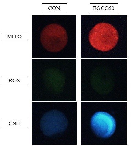
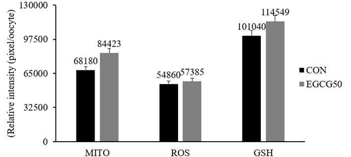
Figures 3: (A) Representative images of ovine oocytes stained with MitoTracker Red®, H2DCFDA® and CellTracker Blue®, respectively, and (B) intracellular levels of mitochondrial activity, ROS and GSH (pixel/oocyte) of oocytes matured in vitro in the absence of antioxidant supplementation (CON) and with 50 µM EGCG (EGCG50). a, b Different letters indicate difference between treatments (P < 0.05)
Cleavage rate
Regarding the cleavage rate, it was possible to observe that the EGCG50 group was superior to the CON group (P-value = 0.03) (Figure 4).
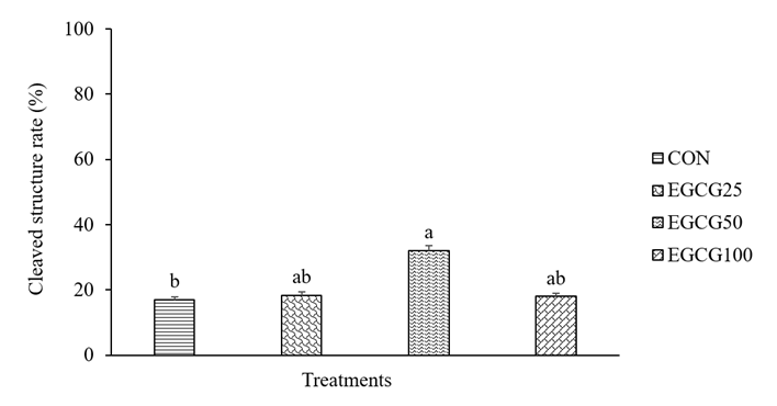
Figure 4: Rate of cleaved structures (%) after in vitro fertilization of matured ovine cells in the absence of antioxidant (CON) or in medium added with 25 µM (EGCG25), 50 µM (EGCG50) and 100 µM (EGCG100) of EGCG. a, b Different letters indicate significant difference between treatments (P < 0.05)
Discussion
The present study showed that supplementation of the IVM medium of ovine oocytes with EGCG increased the levels of active mitochondria and the concentration of GSH, in addition to increasing the rate of cleaved structures.
Oocytes from all groups showed cumulus cell expansion, as well as a higher percentage of oocytes with fully expanded cumulus cells. When EGCG was added to the bovine oocyte IVM medium at the same concentrations as in the present study, the groups containing 25 µM and 50 µM showed a higher percentage of oocytes with grade 3 expansion, i.e., with all cell layers expanded except the corona radiata; and 4, with expansion in all cell layers, including the corona radiata. When evaluating the group containing 100 µM EGCG, only oocytes with grade 3 expansion were observed (Huang et al., 2018). In another study carried out in the same species, supplementation of the IVM medium with 10 µM and 15 µM EGCG increased the percentage of oocytes with expanded cells, compared to the control group and the group using a lower concentration of the substance (5 µM) (Singh et al., 2018). Showing that varying concentrations of the aforementioned antioxidant are capable of promoting the expansion of cumulus cells. In the present study, although not different from the control group, treatments containing EGCG provided a good expansion rate. A possible explanation for this result is the action of EGCG in increasing the expression of genes related to expansion, including hyaluronan synthase 2 (HAS2), tumor necrosis factor alpha-inducible protein 6 (TNFAIP6), pentraxin 3 (PTX3) and prostaglandin (PTGS2), mainly at intermediate concentrations, such as 25 and 50 µM (Huang et al., 2018). Furthermore, the antioxidant action of EGCG may have contributed to these results, eliminating excess ROS in cumulus cells, thus promoting their greater expansion (Fang et al., 2019).
After IVM, the EGCG50 group showed the highest percentage of oocytes in MII (46.4 %) compared to the control group (22.8 %) and other groups containing 25 µM EGCG (25.0 %) and 100 µM EGCG (13.0 %). Similarly, Huang et al. (2018) observed that supplementing the IVM medium of bovine oocytes with the same concentration (50 µM) of EGCG increased the extrusion rate of the first polar body (88.04 %) compared to the control group (76.04 %). In another study, the concentration of 40 µM EGCG protected porcine oocytes that were previously exposed to the agricultural pesticide Diazinon, ensuring meiotic progression and increasing MII rates, when compared to those matured with the pesticide alone (Gai et al., 2022). Studies show that EGCG acts in the maintenance of the cell cycle by modulating the phosphatidylinositol-3-kinase/protein kinase B (PI3K/Akt) cell signaling pathway (Barberino et al., 2020), in addition to stimulating the activity of the enzyme phosphodiesterase 3A (PDE3A), responsible for causing a decrease in the concentration of intra-oocyte cyclic adenosine monophosphate (cAMP), allowing the activation of the MPF and, consequently, promoting meiotic resumption (Pereira et al., 2020). Therefore, it is believed that, in the present study, the concentration of 50 µM EGCG increased the percentage of oocytes in MII through these actions.
In the present study, there was an increase in the levels of active mitochondria for oocytes matured with 50 µM EGCG, compared to the group without antioxidant. Reinforcing these findings, after supplementing the IVM medium of bovine oocytes with the same concentration of EGCG, the authors obtained greater mitochondrial activity and a more homogeneous distribution, compared to the other treatment groups (Huang et al. 2018). Furthermore, the use of 30 µM EGCG in the IVM of oocytes from mice subjected to diabetes mellitus was able to increase the mitochondrial membrane potential, when compared to the control group (Chao et al., 2023). For our results, hypothetized that EGCG may have acted on the expression of PTEN Induced Kinase 1 (PINK1), a gene that encodes proteins responsible for protecting mitochondria from failure during periods of cellular stress, such as in vitro culture, in addition to preserving ATP levels (Zhou et al., 2022), favoring its greater activity.
The use of 50 µM EGCG also increased GSH levels in oocytes. In a study testing the efficacy of EGCG in porcine oocytes subjected to in vitro aging, the authors found that this substance was able to increase GSH levels after 48 hours of culture, compared to the control group (Zhou et al., 2022). In another study, it was observed that after IVM of bovine oocytes in a medium containing green tea polyphenols, there was an increase in GSH levels, which was attributed to the presence of EGCG in the tea composition (Wang et al., 2007). In addition to acting on GSH, EGCG also has the ability to act on other enzymatic antioxidants, such as CAT, SOD, and glutathione peroxidase (GPx), by increasing their gene expression (Meng et al., 2008).
EGCG acts by promoting the activation of Nuclear Factor Erythroid 2-Related Factor 2 (Nrf2) and, consequently, interrupting the interaction between this important transcription protein and the Keap1 protein, thus preventing its degradation and allowing the accumulation of Nrf2 in the cytoplasm for subsequent migration to the cell nucleus, where it comes into contact with the Antioxidant Response Element (ARE) sequences, a specific pathway that plays an important role in the encoding of antioxidant enzyme genes (Zhang et al., 2020). Therefore, it is believed that this entire mechanism was triggered in oocytes matured in the EGCG50 group, resulting in higher concentrations of GSH, which, associated with higher levels of active mitochondria, may have directly contributed to increased oocyte maturation. Despite the increase in the levels of active mitochondria and GSH, the ROS level remained similar between the groups, and a possible explanation is the composition of the IVM medium, where the presence of some substances, such as SFB, which has a direct action against ROS (Biswas & Hyun, 2021), may have masked the action of EGCG, causing it not to show an additional effect.
In addition to the MI and MII stages, another meiotic stage that presented a significant difference was TI. At this stage, oocytes matured with 25 µM EGCG had a lower rate (9.7 %) compared to the EGCG100 group (23.2 %). After supplementing the IVM medium of bovine oocytes with 25 µM green tea polyphenols, a decrease in the embryonic development rate was observed, probably because this concentration was not sufficient to provide the necessary support for the cells to reach telophase I, the phase preceding prophase II and, finally, metaphase II (Wang et al., 2007). Regarding the EGCG100 group, which had a higher TI rate (23.2 %), the concentration may have been able to provide the necessary support for the oocytes to reach this stage, but not to progress further, as demonstrated by the low MII rate in this group. Highlighting this result, Huang et al. (2018) showed that the use of higher concentrations of EGCG, such as 100 µM (76.95 %) and 200 µM (71.57 %) can decrease the MII rate, compared to the concentration of 50 µM (88.04 %). Such results can be explained by the adverse action of EGCG in high concentrations, probably related to pro-oxidant (Zhang et al., 2023) and antiestrogenic (De Amicis et al., 2012) effects.
The present study showed that the use of 50 µM EGCG in the IVM of oocytes provided a higher rate of cleaved structures compared to the control group. Similar results were observed by Huang et al. (2018), who obtained a higher cleavage rate than the control (86.23 % and 76.07 %, respectively) with 50 µM EGCG in the IVM of bovine oocytes. Furthermore, the use of 10 and 15 µM EGCG during IVM promoted an increase in the cleavage rate of bovine oocytes, compared to the group using 5 µM EGCG and the control (Singh et al., 2018). This improvement in embryonic development could be attributed to the antioxidant activity of EGCG via increased intracellular GSH, as previously seen, which may have acted by preventing oocytes undergoing maturation from suffering from the deleterious effects of EO, thus supporting embryonic development effectively.
Conclusion
In conclusion, the supplementation of IVM medium with 50 µM EGCG improved nuclear maturation of ovine oocytes, associated with increased mitochondrial activity and intracellular GSH levels, as well as increasing the initial cleavage rate in embryos.
Acknowledgments
To the Fundação de Amparo à Ciência e Tecnologia de Pernambuco (FACEPE) for awarding the scholarship, process number IBPG-1820-5.05/22.
References
Alfke, J., Kampermann, U., Kalinina, S., & Esselen, M. (2021). Isolation and structural elucidation of dimeric epigallocatechin 3 gallate autoxidation products and their antioxidant capacity. European Food Research Technology, 247, 2961-2975. https://doi.org/10.1007/s00217-021-03846-3
Andrade, K. O., Monte, A. P. O., Silva, R. L. S., Barberino, R. S., Mota, I. M., Santos, G. C. S., Guimarães, V. S., Silva, G. A. L., Teixeira, C. S., & Matos, M. H. T. (2024). Effect of lactose on the in vitro development of sheep secondary follicles. Animal Reproduction Science, 270, 107578. https://doi.org/10.1016/j.anireprosci.2024.107578
Barberino, R. S., Santos, J. M. S., Lins, T. L. B. G., Menezes, V. G., Monte, A. P. O., Gouveia, B. B., Palheta Jr, R. C., & Matos, M. H. T. (2020). Epigallocatechin-3- gallate (EGCG) reduces apoptosis of preantral follicles through the phosphatidylinositol-3-kinase/protein kinase B (PI3K/AKT) signaling pathway after in vitro culture of sheep ovarian tissue. Theriogenology, 155, 25-32. https://doi.org/10.1016/j.theriogenology.2020.05.037
Biswas, D., & Hyun, S. H. (2021). Supplementation of fetal bovine serum increased the quality of in vitro fertilized porcine embryo. Journal of Advenced Veterinary and Animal Research, 8(4), 589-596. https://doi.org/10.5455/javar.2021.h549
Chao, S., Li, L. J., Lu, J., Zhao, S. X., Zhao, M. H., Huang, G. A., Yin, S., Shen, W., Sun, Q. Y., Zhao, Y., Ge, Z. J., & Zhao, L. (2023). Epigallocatechin gallate improves the quality of diabetic oocytes. Biomedicine & Pharmacotherapy, 159, 11426. https://doi.org/10.1016/j.biopha.2023.114267
De Amicis, F., Santoro, M., Guido, C., Russo, A., & Aquila, S. (2012). Epigallocatechin gallate affects survival and metabolism of human sperm. Molecular Nutrition & Food Research, 56, 1655-1664. https://doi.org/10.1002/mnfr.201200190
Du Plessis, S. S., Makker, K., Desai, N. R., & Agarwal, A. (2008). Impact of oxidative stress on IVF. Expert Review of Obstetrics & Gynecology, 3(4), 539-554. https://doi.org/10.1586/17474108.3.4.539
Fang, Y., Zhang, J., Li, Y., Guo, X., Li, J, Zhong, R., & Zhang, X. (2019). Melatonin-induced demethylation of antioxidant genes increases antioxidant capacity through RORα in cumulus cells of prepubertal lambs. Free Radical Biology and Medicine, 131, 173-183. https://doi.org/10.1016/j.freeradbiomed.2018.11.027
Freitas, V. J. F., & Melo, L. M. (2010). In vitro embryo production in small ruminants. Revista Brasileira de Zootecnia, 39(Suppl), 409-413. https://doi.org/10.1590/S1516-35982010001300045
Gai, Y., He, Z. J., Wang, S. G., Wang, N., Ge, Z. J., Huang, G., Shen, W., Yin, S., & Zhao, M. (2022). Epigallocatechin gallate improves meiosis maturation against Diazinon exposure in porcine oocytes. Theriogenology, 196, 1-9. https://doi.org/10.1016/j.theriogenology.2022.09.016
Gaviria, S. M., Herrera, A. L., Betancur, G. R., Urrego, R., & Echeverri-Zuluaga, J. J. (2019). Supplementation with resveratrol during culture improves the quality of in vitro produced bovine embryos. Livestock Science, 221, 139-143. https://doi.org/10.1016/j.livsci.2019.01.025
Gonzales, I. M., Soares, A. T., Gomes, M. G. G., & Sousa, V. H. (2002). Coleta de Sêmen. En Reprodução assistida em caprinos: inseminação artificial (Vol.1, pp. 18-20). Paraíba.
Guerin, P., El Mouatassim, S., & Ménézo, Y. (2001). Oxidative stress and protection against reactive oxygen species in the pre-implantation embryo and its surroundings. Human Reproduction Update, 7(2), 175-189. https://doi.org/10.1093/humupd/7.2.175
Huang, Z., Pang, Y., Hao, H., Du, W., Zhao, X., & Zhu, H. (2018). Effects of epigallocatechin-3- gallate on bovine oocytes matured in vitro. Asian Australasian Journal of Animal, 31, 1420-1430. https://doi.org/10.5713/ajas.17.0880
Marei, W. F., Wathes, D. C., & Fouladi-Nashta, A. A. (2010). Impact of linoleic acid on bovine oocyte maturation and embryo development. Reproduction, 139, 979- 988. https://doi.org/10.1530/REP-09-0503
Marí, M., De Gregorio, E., De Dios, C., Roca-Agujetas, V., Cucarull, B., Tutusaus, A., Morales, A., & Colell, A. (2020). Mitochondrial Glutathione: Recent Insights and Role in Disease. Antioxidants, 9(10), 1-21. https://doi.org/10.3390/antiox9100909
Mateo-Otero, Y., Yeste, M., Damato, A., & Giaretta, E. (2021). Cryopreservation and oxidative stress in porcine oocytes. Research in Veterinary Science, 135, 20-26. https://doi.org/10.1016/j.rvsc.2020.12.024
Meng, Q., Velalar, C. N., & Ruan, R. (2008). Effects of epigallocatechin-3-gallate on mitochondrial integrity and antioxidative enzyme activity in the aging process of human fibroblast. Free Radical Biology and Medicine, 44(6), 1032-1041. https://doi.org/10.1016/j.freeradbiomed.2007.11.023
Pereira, J. L., Curcio, A. G., Barroso, L. M., Mogollón-Waltero, E. M., Gomes, H. F., Maia, R. C., Viana, K. S., Caldas Bussiere, M. C., Marin, D. F. D., & Dias, A. J. B. (2020). Modulation of phosphatidylinositol 3-kinase activity during in vitro oocyte maturation increases the production of bovine blastocysts. Zygote, 28(5), 371-376. https://doi.org/10.1017/S0967199420000209
Singh, L. W., Barua, P. M., Sonowal, J., Nath, K. C., Borah, P., Borah, R. S., Das, A., Mahanta, D., Das, R., & Malla, W. A. (2018). Antioxidant effect of Epigallocatechin gallate on bovine IVM and IVF. Journal of Entomology and Zoology Studies, 6(4), 401-403.
Torres-Osorio, V., Urrego, R., Echeverri-Zuluaga, J. J., & Lópezherrera, A. (2019). Oxidative stress and antioxidant use during in vitro mammal embryo production. Review. Revista Mexicana de Ciencias Pecuarias, 10(2), 433-459. https://www.scielo.org.mx/pdf/rmcp/v10n2/2448-6698-rmcp-10-02-433-en.pdf
Wang, Y., Yang, C., Elsheikh, N. A. H., Li, C., Yang, F., Wang, G., & Li, L. (2019). HO-1 reduces heat stress-induced apoptosis in bovine granulosa cells by suppressing oxidative stress. Aging, 11(15), 5535-5547. https://doi.org/10.18632/aging.102136
Wang, Z. G., Yu, S. D., & Xu, Z. R. (2007). Effect of supplementation of green tea polyphenols on the developmental competence of bovine oocytes in vitro. Brazilian Journal of Medical Biological Research, 40(8), 1079-1085. https://doi.org/10.1590/s0100-879x2007000800008
Zhang, H. H., Su, W., Zhao, R. S., Li, M., Zhao, S. G., Chen, Z. J., & Zhao, H. (2023). Epigallocatechin-3-gallate improves the quality of maternally aged oocytes. Cell Proliferation, 57(4), e13575. https://doi.org/10.1111/cpr.13575
Zhang, Y., Lin, H., Liu, C., Huang, J., & Liu, Z. (2020). A review for physiological activities of EGCG and the role in improving fertility in humans/mammals. Biomedicine & Pharmacotherapy, 127, 110186. https://doi.org/10.1016/j.biopha.2020.110186
Zhou, D., Sun, M., Jiang, W., Li, X., Lee, S., Heo., Niu, Y., Ock, S. A., & Cui, X. (2022). Epigallocatechin-3-gallate protects porcine oocytes against post-ovulatory aging through inhibition of oxidative stress. Aging, 14, 8633-8644. https://doi.org/10.18632/aging.204368
Zhuang, Y., Quan, W., Wang, X., Cheng, Y., & Jiao, Y. (2024). Comprehensive Review of EGCG Modification: Esterification Methods and Their Impacts on Biological Activities. Foods, 13(8), 1232. https://doi.org/10.3390/foods13081232
Author Contributions Statement
1. Conceptualization, 2. Methodology, 3. Data curation, 4. Formal analysis, 5. Writing - original draft, 6. Writing - review and editing, 7. Supervision.
Andreza Mayara Carneiro Lima contributed to 1, 2, 3, 4, 5 and 6. Luana Keally Pimentel de Oliveira to 1, 2, 3 and 6. Érika Karoline de Oliveira Aureliano to 1, 2 and 6. Damaris Raquel Pires dos Santos to 1, 2 and 6. Bruna Moura da Silva to 1, 2 and 6. Luanna Mendes Souza Rodrigues to 2 and 6. Gabriela Cristina da Silva Santos to 2 and 6. Rafael Soares dos Anjos to 2 and 6. Beatriz Cavalcanti de Freitas to 2 and 6. Neilton de Lima Oliveira Junior to 2 and 6. Brena Ferreira de Menezes to 1, 2 and 6. Jennifer da Silva Veloso to 2 and 6. Pedro Humberto Félix de Souza to 4, 6 and 7. Mabel Freitas Cordeiro to 1, 4, 6 and 7. Maria Helena Tavares de Matos to 1, 4, 6 and 7. Edilson Soares Lopes Júnior to 1, 3, 4, 6 and 7.
Data Availability Statement
The dataset supporting the results of this study is not available.
Ethical Approval Statement
All experimental procedures involving animals were approved by the Animal Ethics Committee of the Federal University of Vale do São Francisco (UNIVASF), under protocol number 0001/300823, and were conducted in accordance with national and institutional guidelines for the care and use of animals.
Nota del editor
El editor José Manuel Verdes aprobó este artículo.